What Is a Pedicle Screw System and How Does It Work?
A Pedicle Screw System is a spinal fixation technology designed to stabilize unstable vertebrae and support controlled fusion. It uses screws placed through the pedicles, the strong bony bridges connecting each vertebral body to its posterior elements. Rods then connect the screws, creating a supportive framework along the spine.
The concept sounds straightforward. It is not.
During surgery, the specialist studies imaging, identifies safe screw paths, and considers bone quality, spinal alignment, and nerve locations. The screws must achieve strong purchase without entering the spinal canal or nearby vascular structures. Navigation systems, fluoroscopy, and robotic assistance may improve accuracy, but they do not replace surgical judgment. Small differences in anatomy can change the safest trajectory.
Spine surgeon John Kostuik, MD, is often credited with the reminder, “The goal of spinal surgery is to make the patient better, not the X-ray better.” That principle keeps the Pedicle Screw System focused on patient function, pain relief, and stability rather than hardware appearance alone. Still, the quotation is commonly repeated without one universally verified original source, so readers should treat the attribution carefully.
This guide explains how the system works, why surgeons select it, and what risks require attention. It will also examine screw design, rod connection, imaging guidance, and postoperative healing. Clinical outcomes vary. Patient anatomy matters. The technology is valuable, but it is not automatically the right choice for every spinal condition. An imperfect X-ray can sometimes accompany a meaningful recovery.
What Is a Pedicle Screw System?
What Is a Pedicle Screw System?
A pedicle screw system is an orthopedic implant used to stabilize parts of the spine. It usually includes screws, connecting rods, and locking components. Surgeons place screws through the vertebral pedicles, which are strong bone bridges behind each vertebral body. The rods connect the screws and help maintain alignment during healing. Unlike a single screw, the system creates controlled support across several vertebrae. It may assist with spinal fractures, deformity, instability, or selected fusion procedures.
During surgery, a specialist uses imaging, anatomical landmarks, and careful measurements to guide placement. The screw does not repair every spinal problem. It mainly limits unwanted movement and supports the treated segment. Bone graft may be added when fusion is planned. The hardware remains while bone healing develops, although removal may be considered later. Results depend on bone quality, surgical accuracy, general health, and rehabilitation. Even careful treatment has limitations.
Tips: Ask what problem the system is designed to address. Discuss screw placement, expected mobility, and possible complications with a qualified spine surgeon. Request a plain-language explanation of imaging findings. Share medications, allergies, smoking history, and previous operations. Recovery instructions matter. Lifting too soon can stress healing tissues. Questions are useful. The best system is not universal.
Key Components and Their Roles
What Is a Pedicle Screw System and How Does It Work?
A pedicle screw system stabilizes selected spinal segments during fusion surgery. Its main anchor is the pedicle screw. The surgeon places it through the vertebral pedicle into the stronger rear portion of the vertebra. This position helps the screw resist movement. However, screw placement requires careful planning. Bone quality, spinal anatomy, and the treatment goal all influence the choice.
The screw head, often called a tulip, holds a connecting rod. Rods link several screws and help maintain alignment while fusion develops. A set screw locks each rod inside the tulip. Some systems may include cross-links for added rotational stability. Not every patient needs every component. This detail matters. A construct should support the planned correction without adding unnecessary hardware. Intraoperative imaging or navigation can help confirm the intended trajectory, but these tools do not remove surgical judgment.
Tips: Ask what each component will do in your procedure. Discuss bone density, imaging, and expected movement after surgery. Hardware alone does not create a fusion; bone healing remains essential. It is also reasonable to ask how the surgeon checks screw position. Explanations should be clear, specific, and tailored to your anatomy. Even experienced teams review the plan carefully, because small anatomical differences can change the safest approach.
How Pedicle Screws Are Placed in the Spine
What Is a Pedicle Screw System and How Does It Work?
A pedicle screw system helps stabilize the spine during fusion surgery. Pedicle screws are placed through the strong, narrow pedicles of the vertebrae. They connect to rods, forming a supportive framework. This structure can limit painful movement while the treated bones heal. The exact plan depends on the patient’s anatomy, spinal condition, and surgical goals.
How Pedicle Screws Are Placed in the Spine
The patient usually lies face down under general anesthesia. The surgeon makes a carefully measured incision over the affected spinal levels. Imaging helps identify each pedicle and plan the safest screw path. A small channel is prepared through the pedicle and into the vertebral body. The screw is then advanced gradually, while the team checks its position. Rods are attached afterward, and bone graft may support fusion.
Accuracy matters greatly. A screw placed too close to a nerve, blood vessel, or spinal canal can cause serious problems. Surgeons may use real-time imaging, navigation, or nerve monitoring to reduce these risks. Even with careful planning, anatomy can vary. No operation is perfectly predictable.
Tips: Ask how the surgical team will confirm screw placement. Discuss imaging, expected recovery, and warning signs before surgery. Follow movement restrictions closely, because healing takes time. Pain improvement may be gradual, not immediate.
What Is a Pedicle Screw System and How Does It Work?
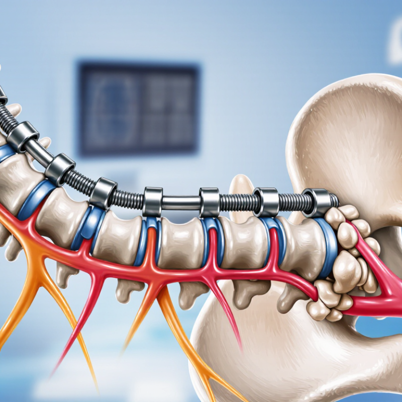
A pedicle screw system stabilizes the spine by anchoring screws through the vertebral pedicles and connecting them with longitudinal rods. In a typical bilateral construct at one vertebral level, two pedicle screws are linked by two rods and secured with set screws.
During placement, the surgeon identifies the pedicle entry point, creates and checks a pilot tract, taps the channel when needed, and inserts the screw along the planned trajectory. Imaging or navigation may be used to confirm alignment and help avoid injury to the spinal canal, nerve roots, and nearby vessels. The exact number of screws, rods, and connectors depends on the spinal levels treated and the surgical plan.
How the System Stabilizes Spinal Segments
What Is a Pedicle Screw System and How Does It Work?
How the System Stabilizes Spinal Segments
A pedicle screw system stabilizes unstable spinal segments from the back. Surgeons place screws through the vertebral pedicles, which are strong bony bridges behind each vertebral body. Connecting rods then link the screws across the affected levels. This structure limits excessive movement while bone healing develops. Think of it as an internal frame, not a replacement spine.
Stability depends on accurate placement, healthy bone, and balanced alignment. Imaging guides the surgeon around the spinal canal and nearby nerves. A 2020 systematic review in the European Spine Journal reported that navigation-assisted techniques generally improve screw-placement accuracy compared with conventional methods. Still, accuracy is not absolute. Small breaches can occur. Clinical judgment remains essential.
The system shares mechanical forces across several vertebrae. It can reduce painful motion during standing, walking, or turning in bed. The North American Spine Society’s clinical guidance emphasizes careful patient selection and radiographic follow-up. Research reviews also report widely varying screw-loosening rates, depending on bone quality, follow-up time, and measurement methods. That matters. A strong construct may still fail in severe osteoporosis, smoking, infection, or poor healing conditions. Surgeons may therefore assess bone density before surgery and recommend follow-up imaging. No implant guarantees fusion.
Common Uses, Benefits, and Potential Risks
What Is a Pedicle Screw System and How Does It Work?
A pedicle screw system stabilizes the spine from the back. Surgeons place screws through the vertebral pedicles, the strong bony bridges behind each vertebra. Connecting rods then hold the segments in a planned position. This structure can support spinal fusion, correct alignment, and protect healing bone.
Common uses include unstable fractures, spinal deformity, spondylolisthesis, and painful degeneration requiring fusion. The main benefit is controlled three-dimensional stability. It may also help restore alignment and allow earlier movement after surgery. However, results depend on diagnosis, bone quality, surgical planning, and rehabilitation. The technology is precise, not magical. A 2020 systematic review in European Spine Journal reported pedicle-screw breach rates near 5%–15%, depending on imaging definitions and surgical methods. A 2017 review in Spine found clinically significant neurological injury was generally below 1% in modern series.
Potential risks include infection, bleeding, nerve irritation, dural injury, screw loosening, rod breakage, and adjacent-segment problems. Revision surgery may be needed. These risks can increase with osteoporosis, smoking, obesity, or complex anatomy.
Tips:
Ask how imaging will guide screw placement. Discuss bone-density testing when appropriate. Confirm the expected fusion levels and recovery limits. Report new weakness, worsening numbness, fever, or wound drainage promptly. Evidence is strong for selected patients, but not every back problem needs instrumentation.