How to Treat Wrist Joint Instability?

Wrist Joint Instability can make ordinary movements feel uncertain. A jar lid turns, but the wrist shifts. A push from a chair brings a sharp ache. Some people notice clicking, weakness, or swelling after an injury. Others have symptoms that build slowly. These signs can overlap with other wrist problems, so symptoms alone cannot confirm the cause.

Hand surgeon William P. Cooney is widely recognized for his work on wrist disorders. A useful summary of the clinical approach reflected in his work is: “Treatment should address the cause of instability, not pain alone.” This is a paraphrase, not a verified verbatim quotation. That distinction matters. Wrist stability depends on small bones, ligaments, and coordinated movement, and damage in one area can change how the whole joint behaves.

Care depends on the injury, its severity, and the person’s daily needs. A clinician may examine wrist motion, grip strength, and tenderness, then use imaging when appropriate. Mild injuries may improve with rest, a brace, and guided rehabilitation. More serious ligament damage may need specialist assessment and, in some cases, surgery. There is no single plan for every wrist. Even a brace can be the wrong fit if it limits needed movement or hides worsening symptoms. An accurate diagnosis is a practical starting point, not a guarantee of a quick recovery. Pain that persists, marked swelling, numbness, or loss of function deserves medical attention. Small details matter.
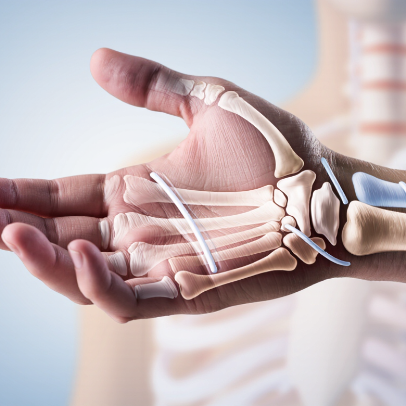
How to Treat Wrist Joint Instability?

Assess Wrist Pain and Laxity; Record Injury Timing and Functional Limits

Wrist instability may feel like the joint shifts, clicks, or gives way when you turn a handle or push up from a chair. Describe where pain sits: near the thumb, little finger, or center of the wrist. Note whether it is sharp, aching, or present only with movement. A clinician can check tenderness, swelling, motion, grip, and ligament laxity, comparing both wrists. Avoid forceful self-testing; it may worsen pain without clarifying the cause. A fall, visible deformity, marked swelling, or new numbness needs prompt medical assessment.

Record when symptoms began and what happened, even if the injury seemed minor. Write down whether pain appeared at once or hours later, and whether it is changing. Small details help. Note trouble lifting a mug, typing, turning a key, or carrying a bag, plus any clicking or episodes of giving way. Include what makes symptoms better or worse, and how long discomfort lasts after activity. Memory is imperfect; a brief daily note can be more useful than a confident guess. Share this record during assessment so the clinician can relate your symptoms and functional limits to examination findings and decide whether imaging or further evaluation is needed.

Confirm Scapholunate Instability: Stress-Radiograph Gap Over 3 mm

Scapholunate instability may cause wrist pain, clicking, or a weak grip after a fall onto an outstretched hand. On a clenched-fist stress radiograph, clinicians measure the space between the scaphoid and lunate bones. A gap greater than 3 mm is commonly considered suspicious for scapholunate widening, according to Andersson and colleagues’ 2017 review in EFORT Open Reviews. The measurement is a useful warning sign. It is not a diagnosis by itself.

Compare the injured wrist with the opposite wrist, and check that positioning is consistent. A poorly centered image or pain that prevents a firm fist can affect the result. Small details matter. The clinician should also consider tenderness, swelling, injury history, and other imaging findings. Some instability appears only under load, while a normal-looking image does not always settle the question. That is an uncomfortable limitation, but worth acknowledging. If symptoms and stress views disagree, a hand specialist may recommend further assessment. The 3 mm threshold helps guide that decision; it should not replace clinical judgment.

How to Treat Wrist Joint Instability? — Confirm Scapholunate Instability

On a stress radiograph, a scapholunate gap greater than 3 mm can indicate scapholunate instability.

Interpretation: A gap over 3 mm is concerning and should be assessed alongside symptoms, examination findings, and comparison with the opposite wrist. A radiographic measurement alone does not confirm a diagnosis.

Classify Ligament Damage and Alignment Before Choosing Treatment

Wrist instability can follow a fall, a forceful twist, or repeated loading. Before choosing treatment, clinicians need to identify which ligament is injured and whether the wrist bones remain aligned. Pain alone cannot answer either question. A small click when turning a door handle may matter, but it does not prove a serious tear.

Assessment combines the injury history, a careful physical examination, and imaging when needed. Plain X-rays can show fractures or changes in bone spacing; special stress views may reveal instability that is not visible at rest. MRI can help assess soft tissues, while CT may clarify complex bone alignment. Findings should be interpreted alongside symptoms, since swelling and guarding can make an examination less clear. The categories are useful, but real wrists do not always fit them neatly.

A mild sprain with stable alignment may improve with temporary support, activity changes, and guided rehabilitation. A substantial ligament tear, persistent widening between bones, or a joint that repeatedly shifts needs prompt specialist assessment. Treatment may involve immobilization or, in selected cases, surgery; the choice depends on the specific ligament, injury timing, function, and overall health. Do not force the wrist through painful tests at home. If instability persists, reassessment matters—even when the first X-ray looked normal.

Begin Stable Injuries with Splinting and Activity Modification

When a clinician confirms that wrist joint instability is mild and stable, initial care often focuses on support and reduced strain. A removable splint can limit painful movement while allowing the fingers to move. It should feel secure, not tight; loosen it if fingers become numb, pale, or unusually cold. Small details matter.

Activity modification helps calm irritated tissues. For a few days, avoid heavy lifting, forceful twisting, and movements that trigger sharp pain, such as opening a stubborn jar. Keep up comfortable tasks, and return to sport or manual work gradually as symptoms improve. Complete rest is not always helpful, and pushing through pain can prolong recovery. That balance can be difficult.

The right splint and wearing schedule depend on the injury, so follow guidance from a healthcare professional rather than copying someone else’s routine. Check the skin daily, especially beneath the edges, and remove the splint as directed for hygiene or prescribed exercises. If pain or instability worsens, swelling increases, or the wrist keeps giving way, arrange reassessment. A splint supports healing; it does not repair every ligament injury.

Refer Acute Complete Tears Promptly; Repair Is Often Considered Within 4–6 Weeks

Wrist joint instability can follow a fall, a hard twist, or a sudden impact. Pain, swelling, a clicking sensation, or difficulty gripping a cup may signal an injury, but symptoms alone cannot show whether a ligament is torn. Acute complete tears need prompt medical assessment. A clinician may examine wrist movement and order imaging to clarify the injury. Do not force the joint to test it.

For some complete tears, surgical repair is considered while the injury is still recent. Repair is often considered within 4–6 weeks, but that is not a guaranteed deadline or a rule for every wrist. The best approach depends on the ligament involved, the degree of instability, and the person’s needs. Delays can make repair more difficult in some cases, so arrange specialist review promptly rather than waiting for pain to settle. Treatment may also include temporary support and guided rehabilitation.

Tips: Note when the injury happened and which movements hurt. Keep the wrist supported as advised, and avoid lifting or twisting until assessed. Small details help. Recovery can be uneven, and a careful reassessment may change the plan.

How to Treat Wrist Joint Instability? – Refer Acute Complete Tears Promptly; Repair Is Often Considered Within 4–6 Weeks

Clinical situation Typical clues Recommended next step Treatment considerations Timing and urgency
Suspected acute complete ligament tear Recent injury with marked pain, swelling, reduced function, or a sense that the wrist is giving way. Examination and imaging are needed to identify the injured structure and severity. Arrange prompt assessment by a hand or wrist specialist. Protect the wrist in a splint while awaiting evaluation. Some complete tears, including selected scapholunate ligament injuries, may be treated with surgical repair. The decision depends on the specific injury, stability, tissue quality, and patient factors. Refer promptly. For selected repairable acute tears, repair is often considered within about 4–6 weeks; this is not a universal deadline or guarantee of surgery.
Possible wrist fracture or dislocation Severe pain or deformity after trauma, substantial swelling, inability to use the hand, or abnormal wrist alignment. Seek urgent medical assessment and imaging. Do not attempt to straighten or manipulate the wrist. Fractures and dislocations can accompany ligament injuries and may require reduction, immobilization, or surgery. Same-day urgent evaluation is appropriate, especially with deformity or severe symptoms.
Persistent pain or instability despite initial care Ongoing clicking, weakness, pain with loading, or recurrent giving-way after an injury. Reassessment is advised. X-rays may be followed by specialist-directed MRI, CT, or other tests; a normal initial X-ray does not rule out every ligament injury. Treatment may include continued immobilization, rehabilitation, or a procedure, depending on the diagnosis and whether the joint is stable. Arrange follow-up rather than relying on symptoms alone to judge healing.
Stable or lower-grade injury Mild-to-moderate symptoms with no demonstrated major instability or displaced fracture. Use clinician-directed protection and follow-up. Avoid painful activities and return to loading gradually. Many stable injuries are managed without surgery, often with temporary splinting and a staged rehabilitation plan. Follow the treating clinician’s instructions; worsening pain, swelling, numbness, or loss of function warrants earlier review.
After repair or immobilization Recovery phase following a procedure or a prescribed period in a cast or splint. Attend scheduled reviews and follow the surgeon’s or therapist’s restrictions on motion and lifting. Rehabilitation is usually gradual. Starting wrist motion or strengthening too early may jeopardize healing; the plan varies by injury and procedure. Return to work, sport, and heavy loading should be guided by clinical recovery, not by a fixed timetable.

Important: This table is general educational information, not a diagnosis or individual treatment plan. Sudden deformity, a cold or pale hand, new numbness, or rapidly worsening pain requires urgent medical attention.