What Are the Top Pedicle Screw Systems for Spinal Fusion?

A Pedicle Screw System can look deceptively simple on a product page: screws, rods, connectors, and instruments arranged in a clean diagram. In surgery, however, fit and handling matter. A surgeon must consider anatomy, bone quality, the planned fusion levels, and whether the system supports the intended alignment. Small differences in screw design, rod options, and instrumentation can affect workflow. They do not guarantee a better outcome.

Orthopedic spine surgeon and researcher Dr. Alexander R. Vaccaro has contributed extensively to spinal fixation research. A concise paraphrase of a principle reflected in that work is: “Choose fixation to suit the patient’s anatomy and the goals of the procedure.” This is a paraphrase, not a verified verbatim quotation. The distinction matters. Product comparisons should rest on published evidence, regulatory information, and experienced clinical judgment—not promotional claims alone.

So, what are the top pedicle screw systems for spinal fusion? There is no universal winner. Surgeons may weigh construct strength, available screw sizes, ease of placement, imaging compatibility, and support for different surgical approaches. Hospital teams also consider training, instrument availability, and consistency across procedures. These details are practical. A tray that is difficult to assemble can disrupt a demanding operation.

The comparison ahead should be useful, but not absolute. Evidence varies by system and patient group. Some questions remain unsettled. That deserves attention.
What Are the Top Pedicle Screw Systems for Spinal Fusion?

Pedicle Screw Systems: Their Role in Lumbar Spinal Fusion

Pedicle screw systems help stabilize the lower spine during lumbar fusion. Screws pass through the pedicles, the strong bridges of bone beside each vertebra, and connect to rods. This structure limits unwanted movement while bone grows across the intended fusion area. The screws do not make the bone fuse by themselves. The hardware is not the whole story. Bone health, surgical technique, and the patient’s recovery all matter.

System design can vary in screw shape, diameter, and adjustment features. A surgeon selects components to fit the patient’s anatomy and the goals of the operation, often using imaging to plan placement. Fit matters. A construct that works well for one person may not suit another. In practice, even careful planning meets uncertainty: bone quality, spinal shape, and healing can differ from what scans suggest. Risks include nerve irritation, infection, loosening, or failure of the bones to unite. Patients should ask their spine specialist why a particular approach is appropriate, what alternatives exist, and how recovery will be monitored. No single system is best for every lumbar fusion.

Core Specifications: Common 5.5–6.0 mm Rods and 4.5–8.5 mm Screws

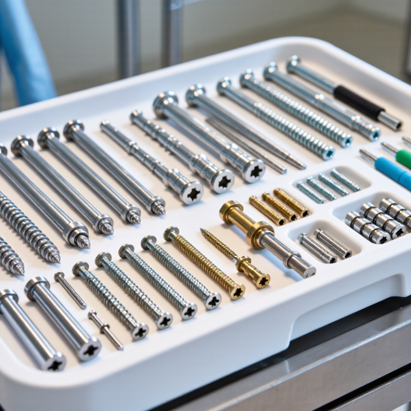
Pedicle screw systems commonly use 5.5 mm or 6.0 mm rods, with screw diameters ranging from about 4.5 mm to 8.5 mm. These are common specifications, not a universal sizing rule. ASTM F1717 tests spinal construct strength in a vertebrectomy model, while ASTM F2193 covers spinal fixation components. Neither standard sets one ideal implant size or predicts an individual patient’s outcome. Fit matters.

Rod and screw dimensions serve different purposes. A rod must match the system’s connectors, while screw diameter and length must suit the planned trajectory and available bone. A larger diameter is not automatically better; anatomy and bone quality matter. These ranges look neat on paper. Real pedicles do not. That is an important limitation when comparing specifications alone.

Tips: Confirm rod-to-connector compatibility, then assess screw dimensions against patient imaging and the surgical plan. Check the current device instructions and relevant ASTM test documentation; a catalog range cannot replace clinical judgment. This detail is easy to overlook.

What Are the Top Pedicle Screw Systems for Spinal Fusion? — Core Specifications: Common 5.5–6.0 mm Rods and 4.5–8.5 mm Screws

System Configuration Common Rod Diameter Common Screw Diameter Typical Screw Lengths Common Design Options Typical Application Considerations
Thoracolumbar posterior fixation 5.5 mm or 6.0 mm Approximately 4.5–8.5 mm Often approximately 25–蠎60 mm, with availability varying by system Polyaxial or monoaxial screw heads; standard or extended-height heads; set-screw rod locking Rod and screw selection depends on spinal level, anatomy, bone quality, and the intended construct.
Small-diameter thoracic fixation Commonly 5.5 mm Often approximately 4.5–6.5 mm Selected to suit the individual vertebra and planned trajectory Low-profile or standard tulip heads; polyaxial options are commonly available Smaller screws may be considered where pedicle dimensions limit available diameter; imaging and anatomical assessment guide selection.
Standard lumbar fixation Commonly 5.5 mm or 6.0 mm Often approximately 6.0–8.5 mm Frequently available in lengths around 35–55 mm Polyaxial or monoaxial heads; screws may be offered with different thread and tip designs Diameter and length are selected according to pedicle size, vertebral anatomy, bone quality, and surgical plan.
Long-segment or higher-load constructs 5.5 mm or 6.0 mm, depending on the system Typically selected from the system’s available 4.5–8.5 mm range Chosen by level and patient anatomy May include stronger rod material options, connectors, and cross-links where compatible Component compatibility is system-specific; rod diameter, material, connectors, and locking components must be verified together.
Revision or challenging rod alignment Usually 5.5 mm or 6.0 mm within the selected system Selected to match the available anatomy and fixation plan Individualized; no single length is appropriate for every level Options may include extended tabs, reduction features, or compatible rod-to-rod connectors Reduction features and connectors are not interchangeable across systems unless compatibility is explicitly specified.
Implant material options Commonly titanium alloy; cobalt-chromium alloy is also used in some systems Commonly titanium alloy; material availability varies Not applicable Material choice may affect stiffness and imaging characteristics Material selection is a clinical and system-specific decision; components should be used only as labeled by the manufacturer.

Note: These are general, non-brand-specific examples of commonly encountered specifications, not a ranking or a purchasing recommendation. Actual dimensions, materials, compatibility, indications, and available options vary by implant system and region. Final component selection should follow the device labeling and the treating surgeon’s assessment.

Leading Platforms: CD HORIZON, SOLERA, CREO, Xia, and EXPEDIUM

The five leading pedicle-screw platform families should be compared by fit and workflow, not by reputation alone. Surgeons assess screw diameter and length options, polyaxial head movement, rod compatibility, and reduction tools. These details matter when a patient’s anatomy is tight or a rod needs careful contouring. Fit matters. No single feature makes a system best for every fusion.

AHRQ’s HCUP Statistical Brief 175 estimated 488,000 inpatient spinal fusion procedures in 2011, a 70% increase from 2001. That figure shows the scale of use, not that one implant platform performs better. Reliable head-to-head evidence can be limited, so claims about superiority deserve scrutiny. That is an uncomfortable gap, but an important one. Compare published clinical outcomes, complication reporting, and the evidence for the specific procedure—not just catalogue features.

Tips: Ask whether the platform offers the screw sizes and connectors needed for the planned anatomy. Check that the surgical team is trained on its instruments, and review outcome data with a clinician. A familiar system can help workflow, but familiarity alone is not proof of better results.

How Surgeons Compare Systems: Screw Options, Navigation, and MIS Access

Surgeons compare pedicle screw systems by how well their components fit a patient’s anatomy and the planned fusion. Screw diameter, length, thread design, and available rod connections all matter. A narrow pedicle may limit the options. Fit matters. The strongest-looking screw is not automatically the right choice; bone quality and safe placement also shape the decision.

Navigation tools can display a planned path over live imaging, while three-dimensional imaging or robotic guidance may help execute that plan. These tools can support accuracy, but they do not replace anatomical judgment or careful registration. A small registration shift can matter. Surgeons also consider image quality, setup time, radiation exposure, and whether the team can verify the trajectory during surgery.

For minimally invasive surgery, systems are compared by their percutaneous instruments, rod-delivery options, and access towers. A compact setup may reduce muscle disruption, yet limited exposure can make adjustments harder. Not always. Instrument feel, visibility, and operating-room workflow deserve attention, too. There is no perfect system for every case, and published accuracy figures may not capture each surgeon’s learning curve or a patient’s specific anatomy.

Clinical Evidence: Reported Fusion Rates Vary, Often Around 80%–95%

Clinical reports often place spinal fusion rates between 80% and 95%, but that range needs context. Studies may count patients or individual spinal levels, use different imaging methods, and follow people for different lengths of time. A rate from one trial cannot be transferred directly to every patient or pedicle screw system. Numbers vary.

Fusion depends on more than the screws. The surgical approach, number of treated levels, bone quality, smoking, and overall health can all influence healing. Researchers may assess fusion with X-rays or CT scans, and those methods do not always show the same details. A solid-looking image also does not guarantee pain relief or restored function.

When comparing clinical evidence, look for clear fusion definitions, adequate follow-up, and patient characteristics similar to yours. Studies with small groups or short follow-up can make results look more certain than they are. It is tempting to rank systems by a single percentage; that is an imperfect shortcut. The reported 80%–95% range describes outcomes across selected studies, not a promise for an individual operation. Discuss how a surgeon measures fusion and weighs the evidence for your specific case.